


painon the back is experienced at least once in a lifetime by 4 out of 5 people. For the working population, they aremost common cause of disabilitywhat determines its social and economic importance in all the countries of the world. Among the diseases that are accompanied by pain in the lumbar spine and extremities, one of the leading places is occupied by osteochondrosis.
Osteochondrosis of the spinal column (OP) is a degenerative-dystrophic lesion of the same, starting from the nucleus pulposus of the intervertebral disc, extending to the fibrous ring and other elements of the medullary segment with a frequent secondary effect on the adjacent neurovascular formations. Under the influence of unfavorable static and dynamic loads, the elastic (gelatinous) nucleus pulposus loses its physiological properties: it dries out and sequesters over time. Under the influence of mechanical loads, the fibrous annulus of the disc, which has lost its elasticity, protrudes, and subsequently fragments of the nucleus pulposus fall through its cracks. This leads to the appearance of acute pain (lumbago), because. peripheral parts of the annulus fibrosus contain receptors for the nerve of Luschka.
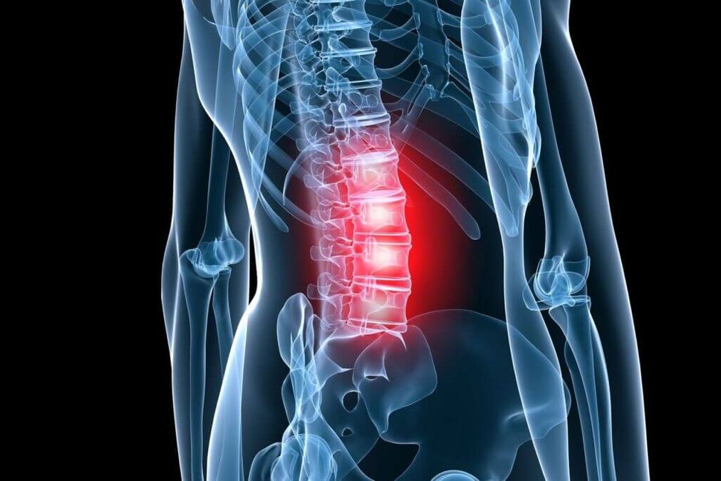
Stages of osteochondrosis
The intradiscal pathological process corresponds to stage 1 (period) (OP) according to the classification proposed by Ya. Yu. Popelyansky and A. I. Osna. In the second period, not only the depreciation capacity is lost, but also the fixation function with the development of hyperlaxity (or instability). In the third period, the formation of a hernia (bulge) of the disc is observed. Depending on the degree of its prolapse, the herniated disc is divided intoelastic overhangwhen there is a uniform protrusion of the intervertebral disc, andkidnapped bump, characterized by the unequal and incomplete rupture of the fibrous ring. The nucleus pulposus moves into these rupture sites, creating local bulges. With a partially prolapsed disc herniation, all layers of the annulus fibrosus are torn, and possibly the posterior longitudinal ligament, but the hernial bulge itself has not yet lost contact with the central part of the nucleus. A completely prolapsed disc herniation means that not its individual fragments, but the entire nucleus, prolapses into the lumen of the spinal canal. According to the diameter of the disc herniation, they are divided into foraminal, posterolateral, paramedian and median. The clinical manifestations of disc herniation are varied, but it is at this stage that various compression syndromes often develop.
Over time, the disease process can move to other parts of the spinal motion segment. An increase in the load on the vertebral bodies leads to the development of subchondral sclerosis (hardening), then the body increases the bearing area due to marginal bone growths around the entire perimeter. Joint overload leads to spondyloarthrosis, which can lead to compression of neurovascular formations in the intervertebral foramen. It is these changes that are noted in the fourth period (stage) (OP), when there is a total lesion of the spinal movement segment.
Any schematization of a disease as complex and clinically diverse as OP is, of course, quite arbitrary. However, it allows to analyze the clinical manifestations in their dependence on morphological changes, which allows not only to make a correct diagnosis, but also to determine specific therapeutic measures.
Depending on which nerve formations the herniated disc, bone growths and other affected structures of the spine have a pathological effect, reflex and compression syndromes are distinguished.
Lumbar osteochondrosis syndromes
Acompressioninclude syndromes in which a root, vessel, or spinal cord is stretched, squeezed, and deformed over the indicated spinal structures. Areflectionthey include syndromes caused by the effect of these structures on the receptors that innervate them, mainly the endings of the recurrent spinal nerves (sinuvertebral nerve of Lushka). Impulses propagating along this nerve from the affected spine travel through the posterior root to the posterior horn of the spinal cord. Switching to the anterior horns, they cause a reflex tension (defense) of the innervated muscles -reflex-tonic disorders.. Switching to the sympathetic centers of the lateral horn from their own or neighboring levels, they cause reflex vasomotor or dystrophic disorders. These neurodystrophic disorders occur mainly in poorly vascularized tissues (tendons, ligaments) at the sites of attachment to bony prominences. Here, the tissues defibrate, swell, become painful, especially when stretched and palpated. In some cases, these neurodystrophic disorders cause pain that occurs not only locally, but also at a distance. In the latter case, the pain is reflected, it seems to "shoot" when touching the diseased area. These zones are called activation zones. Myofascial pain syndromes can occur as part of referred spondylogenic pain.. With prolonged tension of the striated muscle, the microcirculation is disturbed in certain areas of it. Due to hypoxia and edema in the muscle, zones of seals are formed in the form of nodules and strands (as well as in ligaments). The pain in this case is rarely local, it does not coincide with the innervation zone of certain roots. Reflex myotonic syndromes include piriformis syndrome and popliteus syndrome, the characteristics of which are discussed in detail in numerous manuals.
Areflex local pain syndromes (local)in lumbar osteochondrosis, lumbago is attributed to the acute development of the disease and low back pain in the subacute or chronic course. An important circumstance is the established fact thatLow back pain is a consequence of intradiscal displacement of the nucleus pulposus.. As a rule, this is a sharp, often stabbing pain. The patient, as it were, freezes in an awkward position, cannot unfold. An attempt to change the position of the body causes an increase in pain. There is immobility of the entire lumbar region, flattening of the lordosis, sometimes scoliosis develops.
With low back pain - pain, as a rule, pain, aggravated by movement, with axial loads. The lumbar region may be deformed, as in lumbago, but to a lesser extent.
Compression syndromes in lumbar osteochondrosis are also diverse. Among them are distinguished root compression syndrome, caudal syndrome, lumbosacral discogenic myelopathy syndrome.
root compression syndromeoften develops due to a herniated disc at the L levelIV-Lvand mev-Sa, becauseIt is at this level that herniated discs are most likely to develop. Depending on the type of hernia (foraminal, posterolateral, etc. ), one or the other root is affected. As a general rule, one level corresponds to a monoradicular lesion. Clinical manifestations of root compression Lvthey are reduced to the appearance of irritation and prolapse in the corresponding dermatome and to hypofunction phenomena in the corresponding myotome.
paresthesia(numbness, tingling sensation) and shooting pains that spread along the outer surface of the thigh, the front surface of the lower leg to the area of the I finger. Hypoalgesia may then appear in the corresponding area. In muscles innervated by the L rootv, especially in the anterior sections of the lower leg, hypotrophy and weakness develop. First, weakness is detected in the diseased extensor digitorum longus, in the muscle innervated only by the L root.v. Tendon reflexes with an isolated injury to this root remain normal.
By compressing column Sathe phenomena of irritation and loss develop in the corresponding dermatome, extending to the area of the fifth finger. The hypotrophy and weakness mainly affect the posterior muscles of the lower leg. The Achilles reflex decreases or disappears. The patellar reflex is reduced only when the roots of L.two, L3, Lfour. Hypotrophy of the quadriceps, and especially of the gluteal muscles, also occurs in pathology of the caudal lumbar discs. Compressive radicular paresthesia and pain are aggravated by coughing, sneezing. The pain is aggravated by movement in the lower back. There are other clinical symptoms that indicate the development of compression of the roots, their tension. The most commonly tested symptom issymptom of laseguewhen there is a sharp increase in pain in the leg when trying to lift it in an upright state. An unfavorable variant of lumbar vertebrogenic compressive radicular syndromes is compression of the cauda equina, the so-calledcaudal syndrome. In most cases, it develops with prolapsed median large herniated discs, when all the roots at this level are compressed. Topical diagnosis is made in the upper part of the spine. The pains, usually severe, do not extend to one leg, but, as a rule, to both legs, the loss of sensation captures the area of the rider's pants. To the severe variants and the rapid development of the syndrome, sphincter disorders are added. Caudal lumbar myelopathy develops as a result of occlusion of the inferior accessory radiculomedullary artery (often at the root of Lv, ) and is manifested by weakness of the peronial, tibial and gluteal muscle groups, sometimes with segmental sensory disturbances. Often, ischemia develops simultaneously in the segments of the epicon (L5-Sa) and a cone (Stwo-S5) of the spinal cord. In such cases, pelvic disorders also join.
In addition to the main identified clinical and neurological manifestations of lumbar osteochondrosis, there are other symptoms that indicate the defeat of this spine. This is especially clearly manifested in the combination of damage to the intervertebral disc against the background of congenital narrowing of the spinal canal, various anomalies in the development of the spinal column.
Diagnosis of lumbar osteochondrosis
Diagnosis of lumbar osteochondrosisit is based on the clinical picture of the disease and additional examination methods, including conventional radiography of the lumbar spine, computed tomography (CT), CT myelography, magnetic resonance imaging (MRI). With the introduction of spinal magnetic resonance imaging in clinical practice, the diagnosis of lumbar osteochondrosis (OP) has improved significantly. Sagittal and horizontal tomographic sections allow you to see the relationship of the affected intervertebral disc to the surrounding tissues, including an assessment of the lumen of the spinal canal. The size, type of disc herniation, which roots are compressed and by which structures are determined. It is important to establish the conformity of the main clinical syndrome with the level and nature of the injury. As a general rule, a patient with root compression syndrome develops a single root lesion, and the compression of this root is clearly visible on MRI. This is relevant from a surgical point of view, because. this defines operational access.
The disadvantages of MRI include the limitations associated with the examination in patients with claustrophobia, as well as the cost of the study itself. CT is a highly informative diagnostic method, especially in combination with myelography, but it must be remembered that the scan is performed in a horizontal plane, and therefore the level of the alleged lesion must be determined clinically very precisely. Routine radiography is used as a screening test and is required in a hospital setting. On functional imaging, instability is better defined. Various abnormalities of bone development are also clearly visible on spondylograms.
Treatment of lumbar osteochondrosis
With PO, both conservative and surgical treatment is performed. Aconservative treatmentwith osteochondrosis, the following pathological conditions require treatment: orthopedic disorders, pain syndrome, impaired ability to fixate the disc, muscle tonic disorders, circulatory disorders in the roots and spinal cord, nerve conduction disorders, cicatricial adhesive changes, disorders psychosomatic. Conservative treatment methods (CL) include various orthopedic measures (immobilization, spinal traction, manual therapy), physiotherapy (therapeutic massage and physiotherapy, acupuncture, electrotherapy), prescription of drugs. Treatment should be complex, in stages. Each of the CL methods has its own indications and contraindications, but, as a rule, the general isprescription of analgesics, non-steroidal anti-inflammatory drugs(NSAID),muscle relaxantsYphysiotherapy.
The analgesic effect is achieved through the use of diclofenac, paracetamol, tramadol. It has a pronounced analgesic effect.A drugcontaining 100 mg diclofenac sodium.
Gradual (long-term) absorption of diclofenac improves the effectiveness of therapy, prevents possible gastrotoxic effects and makes therapy as convenient as possible for the patient (only 1-2 tablets per day).
If necessary, increase the daily dose of diclofenac to 150 mg, additionally prescribe analgesics in the form of non-long-acting tablets. In milder forms of the disease, when relatively small doses of the drug are sufficient. In the case of predominance of painful symptoms at night or in the morning, it is recommended to take the drug in the evening.
The substance paracetamol is inferior in analgesic activity to other NSAIDs, and therefore a drug was developed, which, along with paracetamol, includes another non-opioid analgesic - propyphenazone, as well as codeine and caffeine. In patients with iscalgia, when caffeine is used, muscle relaxation, decreased anxiety and depression are observed. Good results were observed when using the drug in the clinic to relieve acute pain in myofascial, myotonic and radicular syndromes. According to the researchers, with short-term use, the drug is well tolerated, it practically does not cause side effects.
NSAIDs are the most commonly used drugs for PO. NSAIDs have anti-inflammatory, analgesic, and antipyretic effects associated with the suppression of cyclooxygenase (COX-1 and COX-2), an enzyme that regulates the conversion of arachidonic acid to prostaglandins, prostacyclin, and thromboxane. Treatment should always begin with the appointment of the safest drugs (diclofenac, ketoprofen) at the lowest effective dose (side effects are dose-dependent). In elderly patients and in patients with risk factors for side effects, it is advisable to start treatment with meloxicam and especially with celecoxib or diclofenac/misoprostol. Alternative routes of administration (parenteral, rectal) do not prevent gastroenterological and other side effects. The combination drug diclofenac and misoprostol has certain advantages over standard NSAIDs, reducing the risk of COX-dependent side effects. Furthermore, misoprostol is capable of potentiating the analgesic effect of diclofenac.
To eliminate pain associated with increased muscle tone, it is advisable to include central muscle relaxants in complex therapy:tizanidine2-4 mg 3-4 times a day or tolperisone inside 50-100 mg 3 times a day, or tolperisone intramuscularly 100 mg 2 times a day. The mechanism of action of the drug with these substances is significantly different from the mechanisms of action of other drugs used to reduce the increase in muscle tone. Therefore, it is used in situations where there is no antispastic effect of other drugs (in the so-called unanswered cases). The advantage over other muscle relaxant drugs that are used for the same indications is that with a decrease in muscle tone in the context of the appointment, a decrease in muscle strength does not occur. The drug is an imidazole derivative, its effect is associated with stimulation of a centertwo-adrenergic receptors. Selectively inhibits the polysynaptic component of the stretch reflex, has an independent antinociceptive and mild anti-inflammatory effect. The substance tizanidine acts on spinal and cerebral spasticity, reduces stretch reflexes and painful muscle spasms. It reduces resistance to passive movements, reduces spasms and clonic seizures, and increases the force of voluntary skeletal muscle contractions. It also has a gastroprotective property, which determines its use in combination with NSAIDs. The drug has practically no side effects.
Surgerywith PO, it is carried out with the development of compression syndromes. It should be noted that the presence of the fact of detection of a herniated disc during MRI is not enough for the final decision on the operation. Up to 85% of patients with disc herniation among patients with radicular symptoms after conservative treatment do not undergo surgery. CL, with the exception of a number of situations, should be the first step in helping PO patients. If the CL complex is ineffective (within 2 to 3 weeks), surgical treatment (CL) is indicated in patients with herniated discs and radicular symptoms.
There are emergency indications for PO. These include the development of the caudal syndrome, as a rule, with complete prolapse of the disc into the lumen of the spinal canal, the development of acute radiculomyeloishemia, and a pronounced hyperalgic syndrome, when even opiate blockade does not reduce pain. It should be noted that the absolute size of the herniated disc is not decisive for the final decision on surgery and must be considered in conjunction with the clinical picture, the specific situation observed in the spinal canal according to the tomography (for example, there may be a combination of a small hernia against the background of spinal canal stenosis or vice versa - a hernia is large, but of a medium location against the background of a wide spinal canal).
In 95% of cases with disc herniation, an open access to the spinal canal is used. Several discopuncture techniques have not found wide application to date, although several authors report their efficacy. The operation is performed with both conventional and microsurgical instruments (with optical magnification). During access, removal of bony formations from the vertebra is avoided by primarily using an interlaminar approach. However, with a narrow channel, hypertrophy of the articular processes, fixed median disc herniation, it is advisable to widen access at the expense of bone structures.
The results of surgical treatment largely depend on the experience of the surgeon and the correctness of the indications for a particular operation. According to the apt expression of the famous neurosurgeon J. Brotchi, who has performed more than a thousand operations for osteochondrosis, it is necessary "not to forget that the surgeon must operate on the patient, and not on the tomographic image".
In conclusion, I would like to once again emphasize the need for a full clinical examination and analysis of tomograms in order to make an optimal decision on the choice of treatment tactics for a particular patient.