


The spine is a unique biokinematic system; It is able to withstand loads without damage, but, like any structure, it wears out over time. At a young age, a stable state is maintained thanks to rapid regenerative abilities, but after the age of 50, its supply gradually fades, which leads to the formation of osteochondrosis.
Osteochondrosis is the most common degenerative-dystrophic pathology of the spine, which, as it progresses, spreads to nearby structures of the spinal segment.
Development Theories
The etiology of osteochondrosis is unknown. Currently existing theories about the development of this disease:
- Metabolic.Changes in the metabolism of the vertebral disc due to its dehydration (the amount of water at a young age is 88%, with age the water content decreases to 60%).
- Vascular.Changes in spinal circulation (occurs in adulthood, but earlier development is possible due to injuries, metabolic disorders, infections).
These theories are sometimes combined into one - involution, which is based on a violation of trophism, especially in those tissues in which there are no vessels. In childhood, there is a vascular network in the intervertebral discs, but after the complete formation of the spinal architecture, this network is closed with connective tissue.
- Hormonal theorymore controversial. Hormonal status plays a certain role in the development of osteochondrosis, but it is not appropriate to refer only to hormonal levels. This theory is most relevant for postmenopausal women.
- Mechanical theorytalks about the connection between the appearance of osteochondrosis and the overload of certain parts of the spine.
- Anomaly theory- an isolated case of mechanical theory. Anomalies of the vertebral bodies, fusion of the bodies and lack of fusion of the arch due to inadequate biomechanism stimulate overload of the vertebral discs and lead to the destruction of bone tissue.
These theories have a right to exist, but none of them are universal. It is more correct to call osteochondrosis a multifactorial disease, which is characterized by a genetic predisposition and provoking factors.
Factors that contribute to the development of the disease.
- Gravity factor:For the spine, any non-physiological displacement is nothing more than a trigger for many muscular reactions.
- Dynamic factor: the greater and longer the load on the spine, the longer and longer it is subject to trauma (people prone to forced positions for a long time; constant lifting of heavy objects).
- Dysmetabolic factor:Insufficient nutrition of the spine due to autoimmune disorders, toxic effects.
It is known that the consumption of foods prepared in aluminum dishes causes their accumulation in the bones, which will subsequently contribute to the formation of osteochondrosis. The use of foods made from an alloy of aluminum and iron has an adverse effect on the human body. When preparing food, microparticles enter the gastrointestinal tract and, since they also contain lead, this metal accumulates in the body, the intoxication of which is expressed by neuroosteofibrosis (defective tissue changes at the junction of the tendon and muscle).
- Genetic factor.Each person has an individual level of flexibility, which is directly correlated with the proportion of connective tissue fibers (collagen and elastin) and is genetically inherited. Despite all of the above, there are standards for the proportion of fibers; deviations lead to faster wear of the spine.
- biomechanical factor– non-physiological movements on the articular surface of the spine. This is caused by muscle atrophy (the clinical symptom is pain that occurs when bending and twisting).
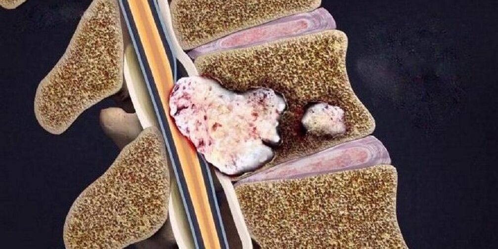
- Aseptic-inflammatory factor– most often a rapid inflammatory process in the intervertebral discs. Microdefects form in the spine due to malnutrition of the spinal disc. In these microdefects, areas of dead tissue form.
Symptoms of osteochondrosis of the spine
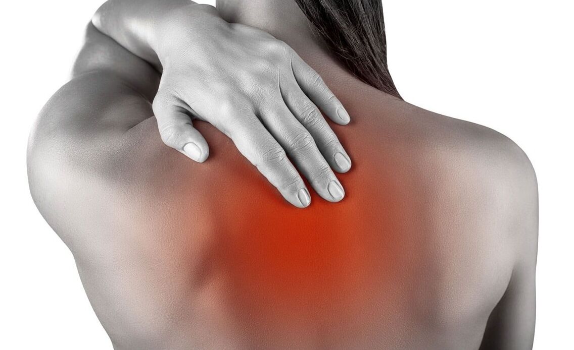
The main symptom of osteochondrosis is back pain, which can be constant or periodic, painful or sharp, and most often intensifies with sudden movements and physical activity.
Osteochondrosis is a common disease among athletes. It arises from a discrepancy between physiological capabilities and motor loads, which contribute to microtrauma and wear of spinal tissue.
The localization of symptoms largely depends on the part of the spine in which the pathological process occurs (cervical, thoracic, lumbosacral). If the pathological process is localized in several parts, this condition is called mixed osteochondrosis.
| Type of osteochondrosis | Cervical | Chest | lumbosacral | Mixed |
|---|---|---|---|---|
| Clinical picture |
|
|
|
The pain is stable or spreads to all parts of the spine. |
| Complications |
|
|
compression myelopathy (compression of the spinal cord by various neoplasms). |
all complications that are possible with cervical, thoracic and lumbosacral osteochondrosis. |
Stages of osteochondrosis
| Stages | First | Second | Third | Four |
|---|---|---|---|---|
| changes in the spine |
|
|
Rupture and displacement of the spinal disc with immersion of other surrounding elements in its cavity, which provokes the development of local symptoms of inflammation. | Destruction of other elements of the intervertebral joint, pathological arrangement of articular surfaces, marginal bone growths. |
| Patient complaints | Absent or indicates discomfort when remaining in the same position for a long time. | Discomfort and pain with certain types of exercise. | Pain in the back, neck, lumbar area, sacrum or coccyx, depending on the location. | Constant pain throughout the spine. |
Differential diagnosis
- Acute myocardial infarction.The pain is concentrated in the heart area and only from there does it radiate (spread) to the neck, lower jaw and arm. The disease begins without reason or after physical activity with the appearance of compressive pain not associated with spinal movement. After half an hour, the pain reaches its maximum, the person develops shortness of breath and fear of death. The diagnosis is confirmed by an electrocardiogram (ECG) and markers of myocardial necrosis.
- Subarachnoid hemorrhage(hemorrhage between the arachnoid and pia mater of the brain). In some cases, due to the toxic effect of spilled blood on the roots of the spine, severe pain in the spine may occur. The main clinical sign is the presence of blood in the cerebrospinal fluid.
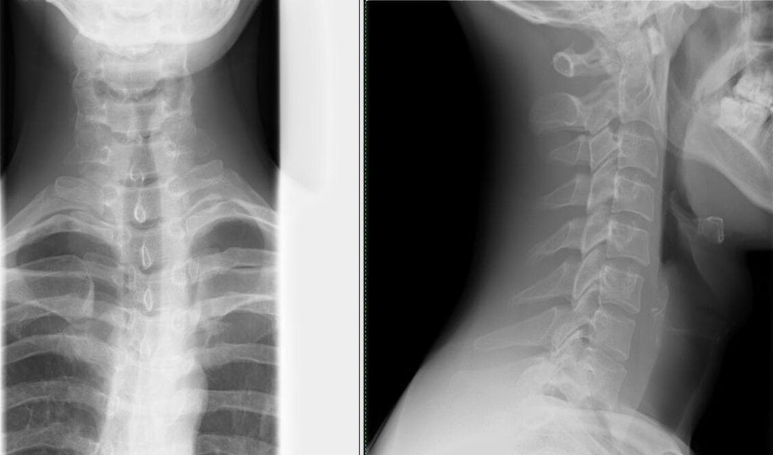
- Spinal anomalies.Minimal exploration: x-ray of the skull and cervical spine in frontal and lateral projections. The most common anomalies of the spine are: fusion of the atlas (the first cervical vertebra) with the occipital bone, depression of the edges of the occipital foramen into the cranial cavity, fusion of the vertebrae, changes in the shape and size of the spine. vertebrae.
- Cervical lymphadenitisIt may also be accompanied by neck pain, sometimes aggravated by bending and twisting. Making a diagnosis is not difficult: enlarged and painful lymph nodes; History of frequent sore throats.
- Multiple myeloma.Pain in the spine appears gradually, against a background of progressive weight loss and periodic fever. The main laboratory sign is protein in the urine.
- Tumor or metastasis in the spine.The evidence in favor of a malignant neoplasm is: progressive loss of body weight, laboratory changes, as well as ultrasound of the sources of metastases: kidneys, lungs, stomach, thyroid gland, prostate.
- Rheumatic and infectious-allergic polyarthritis.differentiated by medical history, moderately elevated body temperature, and predominant damage to large joints.
- Masked depression.Patients "impose" non-existent pathologies (in this context, symptoms of osteochondrosis), an attempt to explain to them the essence of what is happening runs into a wall of misunderstanding. Signs of masked depression are: decreased mood, concentration, and performance; sleep and appetite disturbances; suicidal thoughts and actions.
- Peptic ulcer of the stomach and duodenum, pancreatitis and cholecystitis.They are diagnosed by connecting pain with food intake, laboratory tests (FGDS, general blood test, biochemical blood test, activity of pancreatic enzymes, ultrasound examination of the abdominal organs).
Diagnosis of osteochondrosis.
- Most often, a patient complains to a neurologist, who collects a history of the patient's life and illness and conducts a neurological examination. A neurologist examines the spine in three options (standing, sitting and lying). When examining the back, he pays special attention to the posture, the lower angles of the shoulder blades, the crests of the iliac bones, the position of the shoulder girdle and the expression of the muscles of the back. Deformation, pain and muscle tension are determined during palpation.
- When establishing a diagnosis of osteochondrosis, additional consultation with specialized specialists is necessary to exclude pathologies with similar symptoms (cardiologist, therapist, rheumatologist).
- Carrying out mandatory laboratory tests (general blood test, general urine analysis, biochemical blood test).
- The studies that confirm this are fundamental:
- x-ray of the spine in two projections– the simplest method to identify changes in the spine (narrowing of the gap between the vertebrae);
Depending on the grade, several changes are seen on x-rays:
Degree First Second Third Four x ray signs No radiological signs. Changes in the height of the intervertebral discs. Protrusion (bulging into the spinal canal) of intervertebral discs or even prolapse (loss). Formation of osteophytes (marginal bone growths) at the point of contact of the vertebrae. - computed tomography (CT) and magnetic resonance imaging (MRI)– used not only to identify changes in the spine, but also to determine pathologies in other organs;
- USDG MAG (Ultrasound Dopplerography of the Major Arteries of the Head)– ultrasound examination of the circulatory system of the head and neck, which makes it possible to diagnose the degree of changes in blood vessels as early as possible.
- x-ray of the spine in two projections– the simplest method to identify changes in the spine (narrowing of the gap between the vertebrae);
What treatment methods exist for osteochondrosis?
drug therapyIt must be strictly individual and differentiated, the prescription of medications is made by a doctor after diagnosis.
The main drugs used in the treatment of osteochondrosis:
- Pain relief is carried out with the help of analgesics and non-steroidal anti-inflammatory drugs (NSAIDs). Treatment with NSAIDs should be as short as possible, 5 to 7 days are enough to relieve pain. If the pain is not well controlled and a constant dose of pain-relieving medications is needed, selective COX-2 inhibitors can be taken.
- Antispasmodics reduce pain and relieve muscle spasms.
- Transcutaneous method of pain relief: ointment, the active ingredient of which is an NSAID; anesthetic cream; applications with anti-inflammatory and analgesic medications; Corticosteroids are added for greater effect.
- Treatment aimed at regenerating an inflamed or compressed nerve, as well as improving blood microcirculation: group B vitamins, neuroprotective drugs, nicotinic acid.
- Oral chondroprotectors: glucosamine, chondroitin sulfate. They help stop destructive changes in cartilage when taken regularly. Chondroprotectors are integrated into the structure of cartilage tissue, which increases bone matrix formation and reduces joint destruction. The most favorable composition: chondroitin sulfate + glucosamine sulfate + glucosamine hydrochloride + non-steroidal anti-inflammatory drugs (NSAIDs). These drugs are called combined chondroprotectors.
Non-drug treatment methods:
Neuroorthopedic measures.An important point in the treatment of osteochondrosis is compliance with a rational regimen of physical activity. Staying in bed for a long time and a minimal amount of physical activity not only does not benefit the spine, but also causes a permanent symptom - back pain.
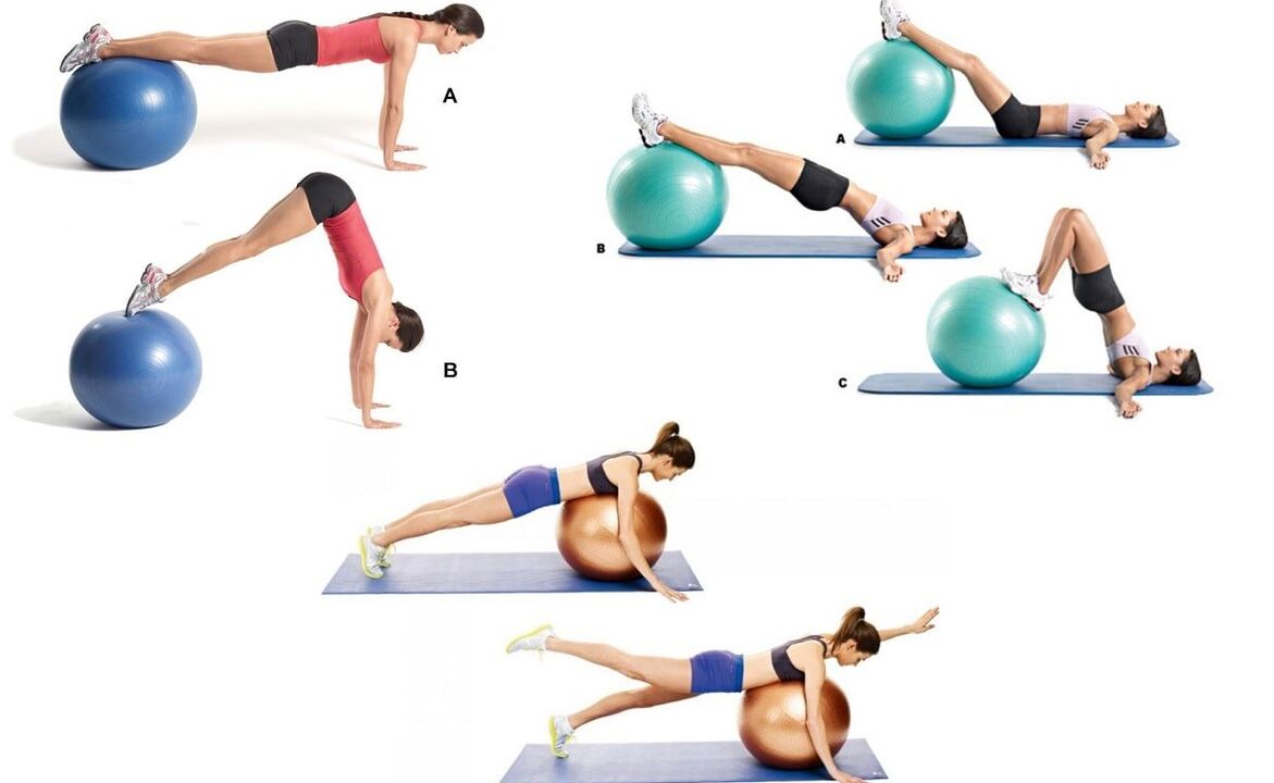
Therapeutic exercise (physiotherapy)It is prescribed when the patient is in satisfactory condition (especially during the period when the signs of the disease decrease), the main goal is to strengthen the muscular corset.
To prevent falls, improve coordination of movements and the functioning of the vestibular apparatus (relevant for elderly patients), balance discs, platforms and paths are used in physiotherapy.
Manual therapywith severe pain in the neck. It is prescribed with special vigilance and according to strict indications. The main goal is to eliminate pathobiomechanical changes in the musculoskeletal system. The main reason for prescribing manual therapy is pathological tension of the paravertebral muscles. Do not forget about a number of contraindications for this type of treatment, which are relevant for osteochondrosis: massive osteophytes (pathological growths on the surface of bone tissue), which are formed at the fourth stage of the development of this pathology.
Physiotherapeutic procedures in the acute period:
- ultrasound;
- phonophoresis;
- ultraviolet irradiation;
- impulsive currents;
- Neuroelectrical stimulation.
Physiotherapeutic procedures in the subacute period:
- electrophoresis;
- magnetotherapy.
Massage.Of all types, a superficial, relaxing massage with rubbing elements is used. As soon as the pain symptom is relieved with the help of massage, they gently move on to more intense rubbing elements. When mastering the technique of acupressure (local) massage, preference is given to this type.
The question of surgical interventions is decided strictly individually, depending on the indications and condition of the patient.
Preventive actions
- Competent selection of furniture (especially in the workplace). The work chair consists of a flat and solid backrest. The bed includes a mattress of moderate hardness, a pillow of medium softness (if possible, an orthopedic mattress and pillow).
- Correction of vision, posture, bite.
- Rational selection of footwear (especially important for drivers). The maximum heel measurement is 5 cm.
- Wear a fixation belt, bandage or corset while working.
- Correction of movements: avoid bending and twisting, lifting weights with a straight back and legs bent at the knees.
- Change your body position more often: do not stand or sit for a long time.
- Proper nutrition: limit the amount of sweet, salty, fatty and spicy foods. The most dangerous food for bones is white sugar, since it separates calcium from bone tissue. The diet should include fruits, berries, vegetables, eggs, nuts, meat, kidneys, liver, fish, legumes and dairy products.
- Protect yourself from sudden changes in temperature, hot water in a bath, sauna, swimming pool, etc. It is especially dangerous, since it relaxes the muscles of the back and even a small injury in this state is not felt, but has tragic consequences for the spine, and even in general for the musculoskeletal system.
- Water procedures are not only a preventive measure, but also a therapeutic one. Swimming combines stretching and relaxation of the muscles.
- Treatment of chronic diseases.
- Active and regular vacations.
Examples of effective exercises to prevent cervical osteochondrosis that can be performed directly at the workplace:
- sitting in a chair, looking forward. The brush covers and supports the lower jaw. Pressing the head forward and down through resistance (tension phase); relaxing and stretching the neck muscles, slowly move your head back (relaxation phase);
- sitting in a chair, looking forward. The right palm is on the right cheek. Slowly tilt your head to the left, try to touch your left shoulder with your ear and stay in this position for 3-5 seconds. Left palm on the left cheek and do the same, respectively, on the right shoulder;
- sitting in a chair, looking forward. Your hands are on your knees. We tilt our head to the right, hold it for 5-7 seconds and very slowly return to the starting position. Then we tilt our head to the left and, accordingly, do the same.
Conclusion
The high frequency and social importance of osteochondrosis determine the scientific interest in this problem. The disease not only affects older people, but is increasingly occurring among young people, attracting the attention of neurologists, neurosurgeons, orthopedic traumatologists and other specialists. Timely diagnosis and adequate treatment of this pathology ensure social adaptation and future quality of life.